Breast cancer is the most common type of female cancer worldwide. Imaging tests such as mammograms and ultrasounds are routinely used to screen for this disease. Next, Flo delves deeper into the subject so you can make a more well-informed decision.
-
Tracking cycle
-
Getting pregnant
-
Pregnancy
-
Help Center
-
Flo for Partners
-
Anonymous Mode
-
Flo app reviews
-
Flo Premium New
-
Secret Chats New
-
Symptom Checker New
-
Your cycle
-
Health 360°
-
Getting pregnant
-
Pregnancy
-
Being a mom
-
LGBTQ+
-
Quizzes
-
Ovulation calculator
-
hCG calculator
-
Pregnancy test calculator
-
Menstrual cycle calculator
-
Period calculator
-
Implantation calculator
-
Pregnancy weeks to months calculator
-
Pregnancy due date calculator
-
IVF and FET due date calculator
-
Due date calculator by ultrasound
-
Medical Affairs
-
Science & Research
-
Pass It On Project New
-
Privacy Portal
-
Press Center
-
Flo Accuracy
-
Careers
-
Contact Us
Screening for Breast Cancer: Mammogram vs. Ultrasound


Every piece of content at Flo Health adheres to the highest editorial standards for language, style, and medical accuracy. To learn what we do to deliver the best health and lifestyle insights to you, check out our content review principles.
Mammogram vs. ultrasound
What is a mammogram?
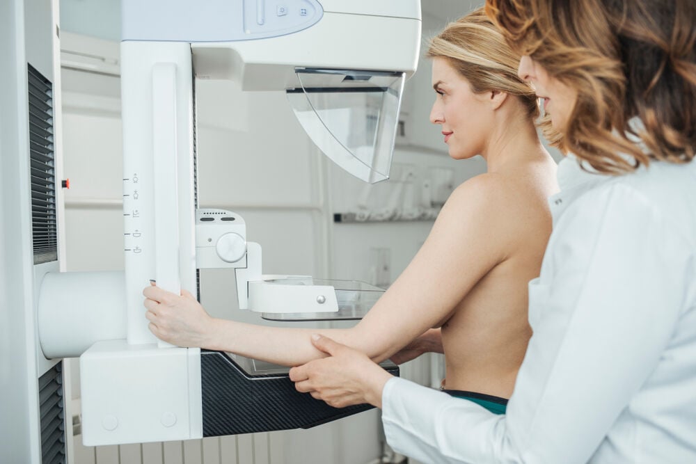
A few key differences separate mammograms and ultrasounds. A mammogram is an x-ray of your breasts used to locate tumors and detect cancer. The mammogram procedure includes the following steps:
- You’ll be asked to undress from the waist up and put on a hospital gown. Depending on the type of equipment, the test may be performed while sitting or standing.
- Your breasts will be placed, one at a time, on a flat surface containing the x-ray plate. A compressor resembling a plastic plate will be momentarily pressed against your breast to help flatten the tissue, making it easier to spot any abnormalities. You’ll likely have to hold your breath as the x-rays are taken from several different angles.
Note that you may feel slightly sore for a few minutes after the mammogram. In some cases, you’ll be asked to return for more x-rays. This isn’t necessarily a cause for concern, it might just mean they need to recheck areas that couldn’t be clearly seen the first time.
What is a breast ultrasound?
A breast ultrasound uses high-frequency sound waves to create an image of your breasts, and the procedure entails the following:
- You’ll be asked to undress from the waist up and put on a hospital gown. While lying on your back on an exam table, your technician will apply gel to your breasts before moving an ultrasound transducer (a wand-like device) over the area.
- The transducer produces sound waves which bounce off your breast tissue and back to the transducer. Waves are then transformed into an image of the inside of your breasts to be examined for any abnormalities.
- You may be asked to raise your arms or turn to the side during the ultrasound to allow for a clearer picture of your breasts.
When should I get a mammogram?

Mammograms are routinely used to check for breast cancer in women who don’t have any signs of the illness. It’s known as a screening mammogram, and involves taking two or more x-rays of each breast. It’s capable of detecting small tumors that cannot be felt during a physical examination. Other abnormalities, such as calcifications which could indicate breast cancer, may also appear during this type of screening.
Diagnostic mammograms, on the other hand, are used when a potential symptom of breast cancer presents itself. Signs include lumps, breast pain, nipple discharge, or changes in breast size or shape, among others. At times, your doctor may order a diagnostic mammogram to further assess the findings of a screening mammogram.
While medical guidelines vary, it’s recommended that women with an average risk of breast cancer begin screening mammograms between the ages of 45 and 54. Women with a greater chance of developing breast cancer might choose to start between the ages of 40 and 49. Be sure to discuss the possible advantages and disadvantages of early breast cancer screening with your doctor.
When should I get a breast ultrasound?
In certain instances, your doctor will suggest an ultrasound instead of a mammogram. Such instances include:
- Onset of potential breast cancer symptoms prior to the age of 25.
- Presence of a breast cyst that must be drained.
- Pregnancy: The use of radiation is one of the key differences between mammograms and ultrasounds. Since ultrasounds don’t rely on radiation, they’re a safer option for mommies-to-be.
- Dense breast tissue: Ultrasounds offer a good alternative when dense tissues prevent proper visualization of your breasts during a mammogram.
How to prepare for a mammogram or ultrasound
As with any other procedure, you can ensure things go as smoothly as possible at your mammogram or ultrasound with a little planning.
How to prepare for a mammogram:
- Try to schedule your mammogram for a time of the month when your breasts won’t be tender. If you’re not menopausal, this usually means the week after your period ends.
- Do not apply deodorant, antiperspirants, lotions, or perfumes before your mammogram appointment. These products contain metallic particles which could alter test results.
- Always bring your prior mammogram findings with you to allow the radiologist to compare new and old images.
How to prepare for a breast ultrasound:
- There’s no need to avoid eating or drinking before a breast ultrasound.
- If you’re asked to fill out a consent form, read it carefully, and don’t be afraid to ask questions.
- Do not apply any lotions or other products on your breasts before your test. Note, the gel used during your ultrasound will not stain your clothes.
- Wear a top that’s easy to remove, like a button-down blouse, to speed up the process of dressing and undressing.
Conclusion: Mammograms vs. ultrasounds
Screening is an important tool when it comes to preventing breast cancer. Interestingly, ultrasounds demonstrate a higher sensitivity for breast cancer in younger women, while mammograms have a higher sensitivity in those older than 60.
While ultrasounds are preferred instead of mammograms for pregnant women, it’s always wise to discuss options with your doctor beforehand. Follow medical recommendations regarding screenings to ensure early diagnosis and treatment of breast cancer, which can have a significant impact on its outcome.


Hey, I'm Anique
I started using Flo app to track my period and ovulation because we wanted to have a baby.


The Flo app helped me learn about my body and spot ovulation signs during our conception journey.


I vividly
remember the day
that we switched
Flo into
Pregnancy Mode — it was
such a special
moment.
Real stories, real results
Learn how the Flo app became an amazing cheerleader for us on our conception journey.

