Perhaps you’ve taken the first step towards conceiving a child through in vitro fertilization, or maybe you’re just interested. If you’d like to see what happens inside the human body during this process, Flo’s got you covered with a detailed IVF timeline.
-
Tracking cycle
-
Getting pregnant
-
Pregnancy
-
Help Center
-
Flo for Partners
-
Anonymous Mode
-
Flo app reviews
-
Flo Premium New
-
Secret Chats New
-
Symptom Checker New
-
Your cycle
-
Health 360°
-
Getting pregnant
-
Pregnancy
-
Being a mom
-
LGBTQ+
-
Quizzes
-
Ovulation calculator
-
hCG calculator
-
Pregnancy test calculator
-
Menstrual cycle calculator
-
Period calculator
-
Implantation calculator
-
Pregnancy weeks to months calculator
-
Pregnancy due date calculator
-
IVF and FET due date calculator
-
Due date calculator by ultrasound
-
Medical Affairs
-
Science & Research
-
Pass It On Project New
-
Privacy Portal
-
Press Center
-
Flo Accuracy
-
Careers
-
Contact Us
IVF Embryo Development Day by Day: Timeline with Description


Every piece of content at Flo Health adheres to the highest editorial standards for language, style, and medical accuracy. To learn what we do to deliver the best health and lifestyle insights to you, check out our content review principles.
Day 0: Pronuclear stage
Fertilization of an egg by a sperm doesn’t happen instantaneously. The two gradually unite over the course of 4–6 hours. The first sign of fertilization is the presence of two round bodies in the egg’s center — the female pronucleus and the male pronucleus. Each contains 23 chromosomes, representing an equal genetic contribution from each parent. In a process known as syngamy, the two cells come together, creating a cell with 46 chromosomes.
Take a quiz
Find out what you can do with our Health Assistant
At this point, the fertilized eggs are checked to ensure everything is going according to plan. Should there be any abnormalities, it’s crucial to catch them before cell division occurs. Even though the embryo would still move through the dividing stages and implant itself in the uterus, it wouldn’t result in a viable pregnancy.
Once the pronuclei join, fertilization is fully completed and a zygote is formed. The entire process — from the moment the sperm and egg connect to the moment cell division begins — takes approximately 30 hours.
Next, the fertilized egg will divide into a two-celled embryo. It continues to duplicate and divide every 10 to 12 hours.
Days 1–3: Cleavage stage
Although the cells are repeatedly dividing during the cleavage stage, the zygote isn’t growing yet. Genetic material is being replicated in each cell, but the overall volume of the zygote remains the same.
The first few days of IVF require constant supervision. As cell division progresses, a culture of fertilized eggs is taken for grading. At this point, zygotes typically have between 6 and 10 cells. The presence of too many cell fragments without any genetic material could indicate a slim chance for viability.
Days 3–5: Morula stage
Once the cleavage stage ends, the morula stage begins. The morula stage prepares the zygote (currently 16 cells, still undergoing division) to grow. The cells arrange themselves into a hollow circle to facilitate transitioning into the blastocyst stage.
Day 5: Blastocyst stage
This is considered the final phase of zygote development. The arrangement described above allows for the formation of two layers of cells. The outer ring provides the tissues needed for a successful pregnancy, including your placenta and amniotic sac. Inside this protective outer ring lies the group of cells that will eventually develop into a baby.
Now, the zygote starts to expand, pushing against its protective shell and preparing itself for implantation. Once the shell is broken, the zygote hatches and adheres to your uterine wall. This is when the zygote develops into an embryo.
Understanding embryo grading
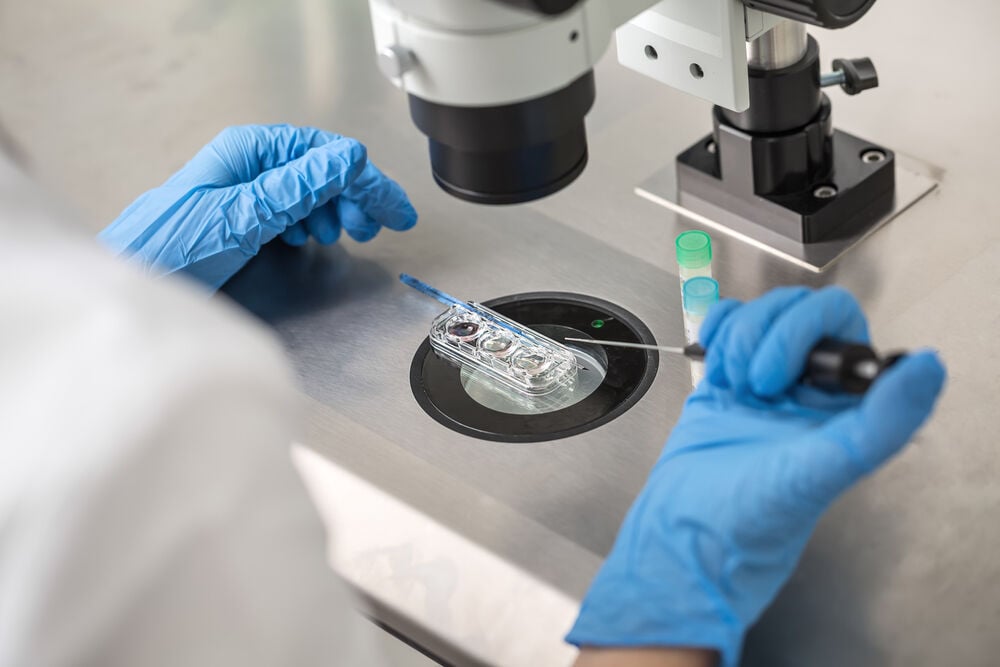
Embryo grading is a tool used by IVF specialists to improve your chances of conception and pregnancy. Your IVF lab will carefully grade your embryos, selecting the ones with the best potential for viable implantation. Grading occurs on both day 3 and day 5, according to different sets of criteria. Implantation is usually done on either day 3 or day 5.
Initially, each fertilized egg is observed to identify the best zygote. Zygotes are evaluated in two ways. The first examines the contact point between nuclear membranes, while the second examines the cell composition of the zygote.
Considered to be of higher quality, grade 1 zygotes contain at least four similarly sized cells, and grade 2 zygotes contain two to four. Grade 3 zygotes, however, have fewer than two blastocysts, which may indicate a lack of viability.
Your IVF lab will carefully grade your embryos, selecting the ones with the best potential for viable implantation. Grading occurs on both day 3 and day 5, according to different sets of criteria.
At the day 3 screening, embryos are observed under a microscope, and the amount of cytoplasm in each cell is measured. Those with a greater number of cells containing genetic material and a good ratio of cytoplasm to nuclei receive a higher grade. Furthermore, cell symmetry is assessed — the more symmetrical a cell is, the better.
By the time day 5 screening begins, the cells are growing, and the zygote is nearly ready to break out of its shell. At this point, a letter grade must be assigned to both the outer ring of cells and the group of cells inside the ring. Both are required for a viable pregnancy.
Grading also considers your age when you’re trying to conceive, fertility history, and the phase of your cycle when selecting the optimal moment for implantation. The ideal number of embryos will most likely be transferred on day 5, and any remaining fully-developed blastocysts may be frozen for future use. Those which haven’t yet achieved a desirable score are cultured for one more day.
Why chromosome screening is important
The decision to conduct chromosome screening is a personal one to consider in advance of IVF. It can only be done when the embryo is outside of your body, during the five-day window between fertilization and implantation.
If either you or your partner have a family history of chromosomal disorders, it’s wise to take screening into consideration. Screening can spot genetic conditions such as muscular dystrophy, sickle cell anemia, cystic fibrosis, and Tay-Sachs.
If you’re using your eggs and donated sperm, screening is particularly useful, since you may not have access to the donor’s full medical history. Screening is also recommended for women over 35 who have had repeated miscarriages, failed IVF treatments, or pregnancies involving chromosomal abnormalities.
If either you or your partner have a family history of chromosomal disorders, it’s wise to take screening into consideration. Screening can spot genetic conditions such as muscular dystrophy, sickle cell anemia, cystic fibrosis, and Tay-Sachs.
Screening occurs on days 5 and 6, after fertilization, and will not affect your chances for viable implantation. The embryo is cryogenically frozen and DNA is extracted for analysis. Chromosome screening is not intended for identifying certain physical characteristics or creating “designer babies.”
The early stages of IVF treatment can be an exciting time for potential parents. Understanding the various phases of embryo development can offer peace of mind during this complex process.
Hey, I'm Anique
I started using Flo app to track my period and ovulation because we wanted to have a baby.
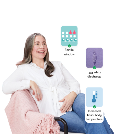

The Flo app helped me learn about my body and spot ovulation signs during our conception journey.
I vividly
remember the day
that we switched
Flo into
Pregnancy Mode — it was
such a special
moment.
Real stories, real results
Learn how the Flo app became an amazing cheerleader for us on our conception journey.


